Fig. 7.
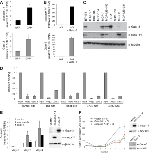
The Gata-3 target gene, caspase-14, promotes differentiation and delays breast tumor formation. (A) Expression analysis of caspase-14 and Gata-3 in GFP+ and GFP− cells from PyMT; MTB; Gata-3+/Tg mammary tumors (means ± standard errors of the mean [SEM] of results for seven tumors per group). (B) Expression analysis of caspase-14 and Gata-3 in immortalized mammary epithelial cells isolated from Gata-3f/f mice, transduced sequentially with pMSCV-cre retrovirus and then Gata-3-expressing (or empty) pBABE retrovirus. (C) Western blot showing the expression of GATA-3, caspase-14, and tubulin in human breast cancer cell lines. (D) ChIP analysis of endogenous GATA-3 binding to four putative GATA-3 binding sites identified within a 10-kb upstream regulatory region of caspase-14 (−484, −3,060, −3,772, and −4,392 from the transcription start site) and a flanking region of caspase-14 promoter with no GATA-3 binding site (control) in MCF-7 cells. Unprecipitated chromatin provided the input control (mean ± SEM of results of three experiments). Mouse IgG and Gata-3 ChIP values for each region were compared using the Student t test: −484 (P = 0.013), −3,060 (P = 0.058), −3,772 (P = 0.028), −4,392 (P = 0.067), control (P = 0.195). (E) β-casein mRNA expression in HC11 cells transduced with control (empty pFU-TA-GFP), pFU-TA-GFP-Gata-3-, or caspase-14-expressing lentiviruses (means ± SEM of results of four independent experiments). Right panel shows Western blot analysis of Gata-3, caspase-14, and β-actin in transduced HC11 cells. (F) Kinetics of tumor formation after orthotopic transplantation of 500,000 MDA-MB-231Luci cells transduced with either pFU-TA-GFP-Gata-3- or caspase-14-expressing lentiviruses or control (vector) virus (means ± SEM of results for six animals per group). Right panel shows Western blot analysis of caspase-14, GATA-3, GAPDH, and tubulin expression in transduced MDA-MB-231Luci cells at the time of transplantation.

