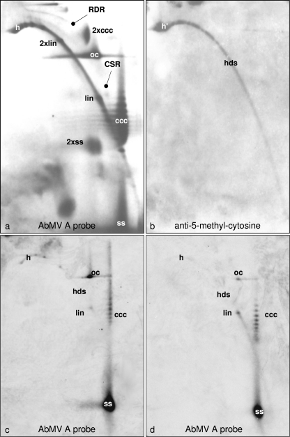
Fig. 6.
Positive detection of methylated viral DNA after 2D agarose gel electrophoresis. Following Southern blotting, either AbMV DNA was probed with full-length DNA A (a) or methylated DNA was visualized with m5C antibody (b). In an independent experiment, viral DNA (100 ng total nucleic acid from AbMV-infected N. benthamiana plants harvested at 21 dpi) was kept untreated (c) or treated with McrBC (d) and then analyzed by 2D agarose gel electrophoresis (first dimension, 0.3% SDS; second dimension, 20 μg/ml chloroquine; 19 h at 45 V). Southern blots were hybridized (c and d) with a probe based on AbMV DNA A lacking the intergenic region. The most prominent viral DNA forms are as described in the legend to Fig. 5 and also included heterogeneous linear dsDNA (h and h′), recombination-dependent replication intermediates (RDR), complementary strand replication intermediates (CSR), and dimeric forms (2×).

