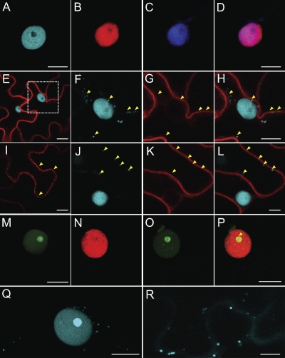
Fig. 6.
Subcellular localization of Bean dwarf mosaic virus (BDMV) movement proteins (NSP and MP) and histone H3 in epidermal cells of N. benthamiana leaves following delivery and expression via Agrobacterium tumefaciens-mediated transient expression. (A) The nucleus of an epidermal cell in leaves infiltrated with H3-ECFP, exhibiting strong fluorescent signal over the nucleoplasm, but not over the nucleolus. (B) As in panel A but with H3-mRFP. (C) A representative nucleus revealed by DAPI staining. (D) Merged images of panels B and C, confirming localization of H3-mRFP over the nucleoplasm. (E) Coexpression of MP-mRFP and H3-ECFP. (F) Magnification of boxed area in panel E, showing egress of H3-ECFP from the nucleus into the cytoplasm in the presence of MP-mRFP; arrowheads identify regions of H3-ECFP accumulation. (G) Localization of MP-mRFP to the nuclear and cellular peripheries (arrowheads). (H) Merged images of panels F and G; arrowheads identify regions of H3-ECFP accumulation outside the nucleus. (I) MP-mRFP localized to punctate structures (arrowheads) either along or over the cell wall and that were subsequently identified as plasmodesmata (see Fig. S7 in the supplemental material). (J) H3-ECFP localized over the nucleus and punctate structures either along or over the cell wall (arrowheads). (K) MP-mRFP localized to cytoplasmic strands (arrowheads) extending from the nuclear to cell periphery. (L) Merged images of panels J and K; arrowheads indicate regions of H3-ECFP accumulation along the cell periphery. (M) NSP-EGFP strongly localized over the nucleolus. (N) H3-mRFP localized over the nucleus and nucleolus in coexpression experiments with NSP-EGFP. (O) Accumulation of NSP-EGFP within the nucleolus and foci in the nucleoplasm in coexpression experiments with H3-mRFP. (P) Merged images of panels N and O; note the colocalization of NSP-EGFP and H3-mRFP over a ring-like structure in or around the nucleolus (arrowhead). (Q) Accumulation of H3-ECFP within the nucleoli of cells infected with BDMV DNA-A and DNA-B. Note also the presence of an H3-ECFP signal located within the cytoplasm. (R) As in panel Q but at a later stage in the BDMV infection process; note the continued egress of H3-ECFP into the cytoplasm and cell periphery, including over PD-like punctate bodies in the cell wall. Scale bars = 10 μm. All CLSM images were collected 48 h after infiltration, except those in panels M to P, which were taken 24 h postinfiltration.

