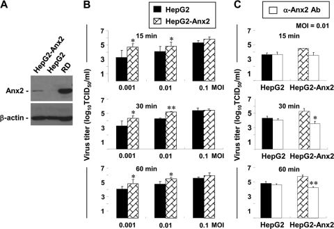
Fig. 7.
Enhanced infectivity of EV71 in HepG2 cells with Anx2 expression (HepG2-Anx2). (A) Western blot analysis of Anx2 expression in HepG2-Anx2 cells. A total-cell lysate (20 μg of protein) from each cell line was resolved by SDS-PAGE and immunoblotting using antibodies against Anx2 and β-actin. (B) Virus yields in HepG2 and HepG2-Anx2 cells. EV71 at each infective dose (MOI, 0.001, 0.01, or 0.1) was allowed to adsorb to 1 × 10⁵ cells for 15, 30, or 60 min, and unbound virus was then washed away. The virus yield at 24 h postinfection was determined by a TCID50 assay with 0.5 log10 serial dilutions of virus. Results are means ± SE for three experiments. Asterisks indicate significant differences from values for HepG2 cells (*, P < 0.05; **, P < 0.01). (C) Blocking effect of the anti-Anx2 antibody in HepG2 and HepG2-Anx2 cells. Cells (1 × 10⁵) were pretreated with an anti-Anx2 antibody or a mouse IgG1 isotype control antibody at 20 μg/ml for 1 h at 37°C, infected with EV71 (MOI, 0. 01) for either 15, 30, or 60 min, washed to remove unbound virus, and cultured in 2% FBS-MEM. The virus yields at 24 h postinfection were determined by a TCID50 assay based on 0.5 log10 serial dilutions of virus. Results are means ± SE for three experiments. Asterisks indicate significant differences from values for control antibody-pretreated cells (*, P < 0.05; **, P < 0.01).

