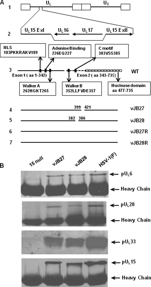
Fig. 1.
Recombinant viruses and coimmunoprecipitation of terminase and portal components. (A) Schematic representations of sequence arrangements of HSV-1 (F) and recombinant viruses relevant to this report. Line 1, HSV-1 (F) DNA. The repeats flanking the unique long (UL) and unique short (US) sequences are indicated by open rectangles. Line 2, gene cluster including UL15 exon I and exon II, UL16, and UL17. Line 3, representation of wild-type pUL15 domains. The solid line represents exon I, and the dashed line represents exon II. Functional domains are indicated in the boxes. Line 4, vJB27 DNA lacking UL15 codons 400 to 420. Line 5, vJB28 DNA lacking UL15 codons 383 to 385. Lines 6 and 7, diagram of UL15 genes from viruses designated vJB27R and vJB28R that were genetically derived from vJB27 and vJB28, respectively, but bear restored UL15 genes. (B) CV1 cells were infected with 15 null, HSV-1(F), mutant virus vJB27 or vJB28 at an MOI of 5 PFU/cell. At 18 h after infection, the cells were washed with cold PBS, lysed in RIPA buffer containing 1 M NaCl2, and immunoprecipitated with anti-pUL15 N antibodies. The immunoprecipitated proteins were eluted in SDS sample buffer, separated on a denaturing 12% polyacrylamide gel, and transferred to a nitrocellulose membrane. The transferred proteins were probed with anti-pUL6, pUL28, pUL33, and pUL15 antibodies as indicated to the right of the figure. The dark band at the bottom of panels A, B, and D is from the IgG heavy chain.

