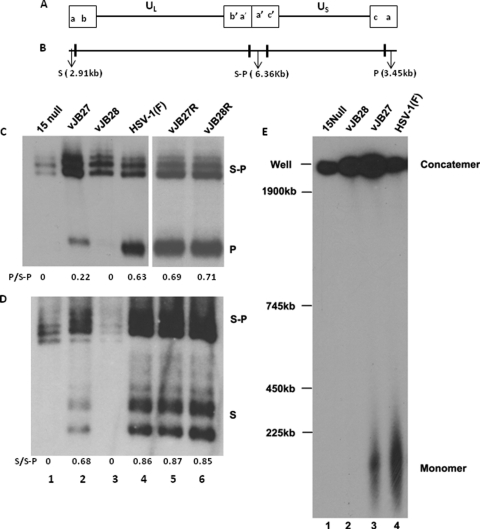
Fig. 2.
Analysis of viral DNA. (A and B) Schematic diagram of the HSV-1 genome showing the positions of the BamHI P or S or S-P joint fragment in HSV-1 DNA. (C) CV1 cells were infected with the indicated viruses and lysed at 15 h after infection, and DNA was purified, digested with BamHI, electrophoretically separated, transferred to a nylon membrane, and probed with radiolabeled DNA representing the terminus of the short component of the viral genome (BamHI P). Fluorographic images captured on X-ray film are shown. Bands were quantified by densitometry, and the ratios of P to S/P fragments are shown below each lane. The positions of the terminal BamHI P fragment and junction fragment S-P are also shown. (D) A different blot from that shown in panel C was hybridized with radiolabeled DNA derived from the long component terminus (BamHI S). The ratios of the S to S-P fragments are shown below each lane. (E) Southern blot analysis of viral DNA separated by pulsed-field gel electrophoresis. CV1 cells were infected with the indicated virus and, at 15 h after infection, were placed in agarose plugs as described in Materials and Methods. After pulsed-field gel electrophoresis, the DNA was denatured, transferred onto a nylon membrane, and probed with 32P-labeled BamHI P fragment of HSV-1 genome. The positions of size standards are indicated to the left.

