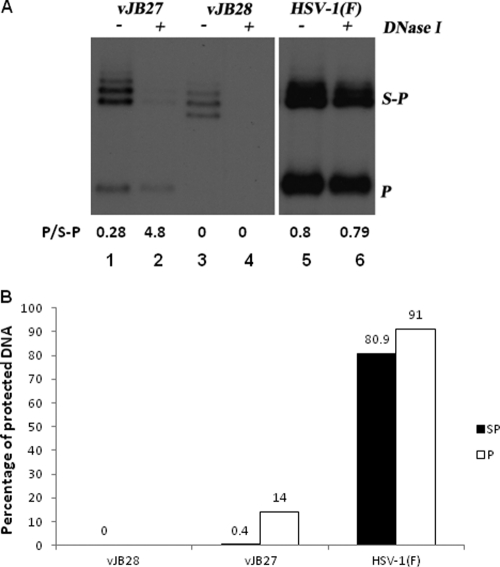
Fig. 4.
DNase protection assay. (A) Fluorographic image of HSV-1 DNA probed with BamHI P. CV1 cells were infected with HSV-1(F) or UL15 mutants at an MOI of 5 PFU/cell. Cells were lysed at 18 h and were incubated in the presence or absence of DNase I. Nuclease treatment was stopped by treatment with proteinase K, and viral DNA was purified and analyzed by Southern blotting as described in the legend to Fig. 2. The blot was analyzed in a Molecular Dynamics Storm 860 PhosphorImager, followed by quantification with ImageQuant software. The ratios of the amounts of BamHI P to S-P fragments are shown below each lane. (B) The percentages of S-P (filled) and P-specific (empty) radioactivity protected from DNase I digestion in the different viral DNAs (i.e., the ratio of the activity of the corresponding bands in treated versus untreated samples) are indicated.

