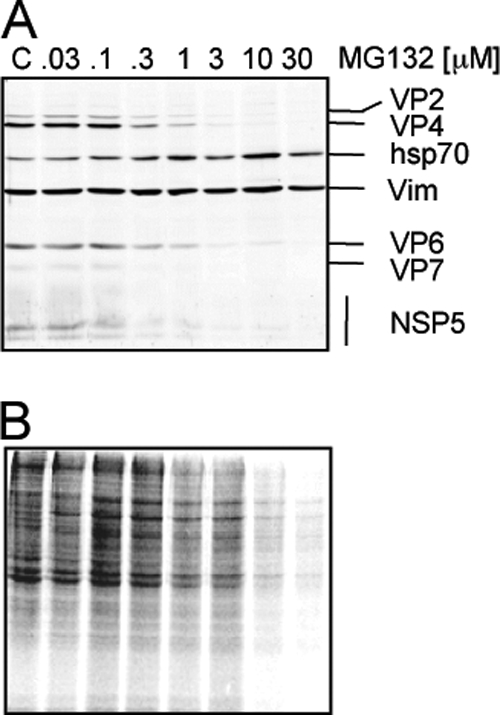
Fig. 2.

Inhibition of proteasome activity reduces viral and cellular protein synthesis. Cells grown to confluence in 96-well plates were infected (A) or not (B) with rotavirus RRV at an MOI of 3 and then were incubated with the indicated concentrations of MG132 for 8 h, and the cell monolayer was solubilized in Laemmli sample buffer. (A) The proteins were analyzed by Western blotting for detection of rotavirus structural proteins and the virus nonstructural protein NSP5 using specific antibodies. Vimentin (Vim) was used as a loading control, and Hsp70 was detected as a stress indicator. (B) Uninfected MA104 cells were incubated for 8 h with the indicated concentration of MG132. During the last hour of incubation 25 μCi/ml of Easy Tag EXPRESS-35S labeling mix was added; after this period, the cells were washed and lysed with Laemmli sample buffer. Proteins were collected and resolved by SDS-PAGE and then subjected to autoradiography.
