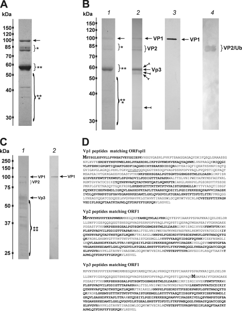
Fig. 4.
Characterization of BgDNV structural proteins. (A) SDS-PAGE of BgDNV structural proteins obtained by infection of B. germanica adults and purification in a CsCl gradient. Protein molecular mass markers are indicated on the left in kilodaltons. The protein bands observed with Coomassie brilliant blue R250 are indicated as follows: ∼100-kDa band, arrow; ∼85-kDa and ∼80-kDa bands, small bracket and one asterisk; ∼57-kDa and ∼56-kDa bands, small bracket with two asterisks; ∼41-kDa, 36-kDa, and 27-kDa bands, large bracket with three asterisks. (B) Western blot analysis of BgDNV capsid proteins. In lane 1, BgDNV capsid proteins were resolved by SDS-PAGE, blotted onto PVDF membrane, and then stained with Coomassie brilliant blue R250. All of the detected bands are indicated as in panel A. In lane 2, the same blot was probed with antibodies to the common C terminus of the capsid proteins. The ∼100-kDa band is indicated by an arrow and designated VP1, the ∼85-kDa and ∼80-kDa bands are indicated by a bracket and designated VP2, the ∼57-kDa band is indicated by an arrow and designated VP3, and the ∼36-kDa band and several bands near VP3 are indicated by feathered arrows. In lane 3, the same blot as in lane 1 was probed with antibody to the unique VP1 N terminus. In lane 4, detection of ubiquitin conjugation with VP2 capsid protein. The same blot as in lane 1 was probed with monoclonal antibody to ubiquitin (P4G7 clone; Covance). Two bands corresponding to VP2 are indicated by a bracket. Protein molecular mass markers are indicated in kilodaltons on the left. (C) SDS-PAGE of BgDNV capsid proteins in BGE-2 cells. Protein extracts were obtained from BGE-2 cells infected with BgDNV, subjected to 10% SDS-PAGE, and blotted onto PVDF membranes. Lane 1, immunoreactivity with antibodies to the common C terminus. Lane 2, immunoreactivity with antibodies to the unique VP1 N terminus. BgDNV capsid proteins are indicated as in panel B, lane 2. Unique BgDNV capsid proteins (presumably resulting from intracellular protease digestion of the native capsid proteins) are indicated by a large bracket and four asterisks. Protein molecular mass markers are indicated in kilodaltons. (D) Examples of mass spectroscopy-detected peptides for virus proteins VP1, VP2, and VP3 matching the corresponding BgDNV ORFs. Matched peptides are shown in bold. The methionine residue (M) driving the synthesis of VP1, VP2, and VP3 is shown in larger bold font. The amino acid sequence where two ORFs are joined during a splicing event to produce ORFspl is underlined. The G shown in italics in ORFspl originates as a consequence of the junction between splice donor and acceptor sites.

