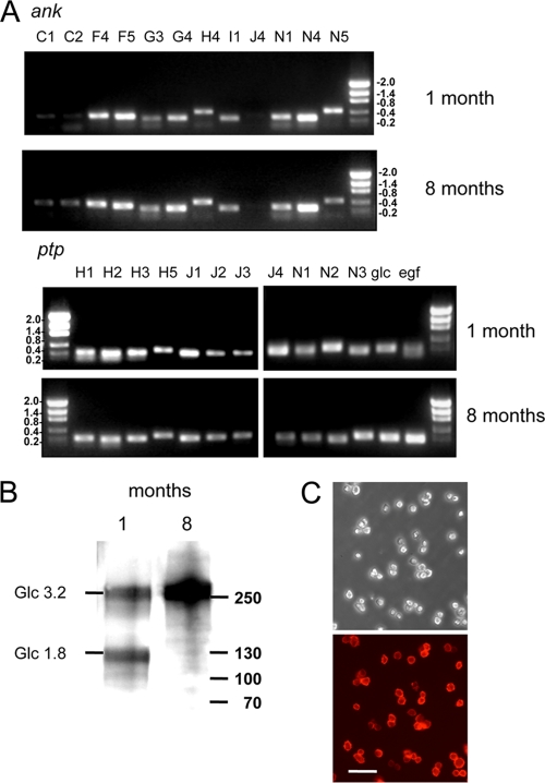
Fig. 2.
MdBV transcripts are persistently detected in infected CiE1 cells. (A) Total RNA was isolated from CiE1 cells at 1 and 8 months postinfection, followed by RT-PCR analysis using primers specific for selected ank and ptp gene family members. Primers that amplify both glc1.8/3.2 and egf1.0/1.5 were also used. Amplicons for most genes were detected at both 1 and 8 months postinfection. Size markers (kb) are indicated to the right or left of the gels. (B) Immunoblot showing the presence of Glc1.8 and Glc3.2 in CiE1 cell extracts 1 month postinfection and the presence of only Glc3.2 in cells at 8 months postinfection. Molecular mass markers (in kDa) are indicated at the right. (C) Phase-contrast (top) and epifluorescence (bottom) micrographs of CiE1 cells at 8 months postinfection labeled with anti-Glc1.8/3.2 and visualized by using an Alexa 564 secondary antibody. The scale bar in the bottom image equals 180 μm.

