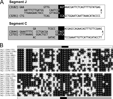
Fig. 8.
MdBV genomic DNAs contain similar host integration motifs (HIMs). (A) Predicted stem-loop structures for the segment J and C HIMs generated by Mfold. Gray highlights nucleotides that form the base of the stem, black highlights the tetramers that identify the boundary site of integration of each segment into host cells, and white highlights the predicted loop domain that is deleted with integration into the host genome. (B) Sequence alignment of the HIMs from selected MdBV genomic segments. The position of the motif on each segment is indicated at the left. Identical nucleotides are indicated in black. The gray, black, and white lines above and below the alignment correspond to the stem and loop regions shown in panel A.

