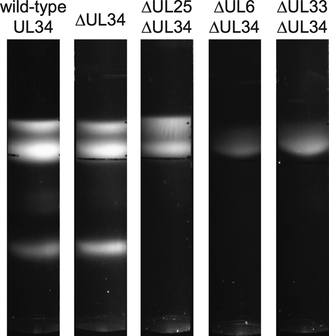
Fig. 2.

Sedimentation of PRV capsids through sucrose. Intranuclear capsids recovered from cells infected with viruses encoding a myc-pUL31 fusion were separated on continuous 20 to 50% sucrose gradients. Three light-diffracting bands are visible for the viruses with either a wild-type UL34 allele or a UL34 deletion allele. Only A and B capsid bands are apparent in the absence of pUL25, and only B capsids are visible in the absence of either pUL6 or pUL33.
