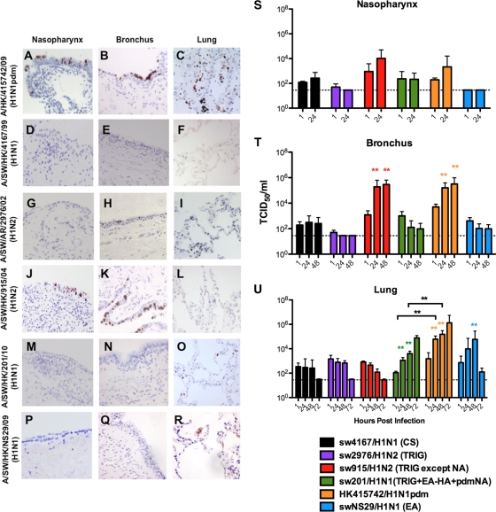
Fig. 1.
Expression of influenza virus nucleoprotein (reddish brown) in the upper, conducting, and lower respiratory tract (A, D, G, J, M, and P), in the nasopharynx (B, E, H, K, N, and Q), and in the bronchi and lungs (C, F, I, L, O, and R) infected with influenza virus A/HK/415742/09 (H1N1pdm), A/SW/HK/4167/99 (H1N1), A/SW/AR/2976/02 (H1N2), A/SW/HK/915/04 (H1N2), and A/SW/HK/201/10 (H1N1) at 48 h postinfection (hpi). Viral replication kinetics in ex vivo cultures of nasopharynx (S), bronchi (T), and lung biopsy specimens (U) infected with 106 TCID50 of influenza viruses/ml by virus titration at 37°C were also evaluated. The chart shows the means and standard errors of the means of the virus titer pooled from at least three independent experiments. Horizontal dotted line denotes the detection limit of the viral titration assay. Colored asterisks indicate the statistically significant increases in viral yield compared to 1 hpi, and black asterisks indicate statistically significant differences between HK415742/H1N1pdm and sw201/H1N1 in figure U. **, P < 0.005.

