Fig. 3.
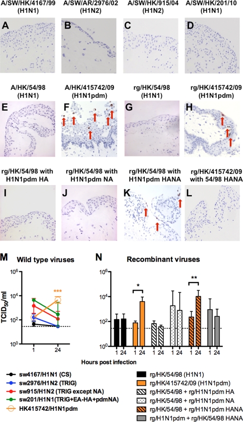
Ex vivo organ cultures of conjunctiva infected with wild-type influenza A/SW/HK/4167/99 (H1N1) (A), A/SW/AR/2976/02 (H1N2) (B), A/SW/HK/915/04 (H1N2) (C), A/SW/HK/201/10 (H1N1) (D), A/HK/54/98 (H1N1) (E), and A/HK/415742/09 (H1N1pdm) (F) viruses and the recombinant influenza viruses rg/HK/54/98 (H1N1) (G), rg/HK/415742/09 (H1N1pdm) (F), rg/HK/54/98 with the HA of A/HK/415742/09 (H1N1pdm) (I), rg/HK/54/98 with the NA of A/HK/415742/09 (H1N1pdm) (J), rg/HK/54/98 with the HANA of A/HK/415742/09 (H1N1pdm) (K), and rg/HK/415742/09 (H1N1pdm) with the HANA of A/HK/54/98 (H1N1) (L) at 24 hpi and stained for influenza virus A nucleoprotein in reddish brown, as indicated by arrows. (M and N) Wild-type (M) and recombinant (N) influenza virus yield at 24 hpi after infection with 106 TCID50 of the virus/ml at 33°C. The chart shows the means and the standard errors of the virus titers pooled from three independent experiments. Horizontal dotted line denotes detection limit of the viral titration. *, P < 0.05; **, P < 0.005; ***, P < 0.0005.

