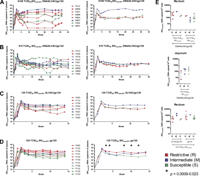
Fig. 4.
SIVmac251 viral and proviral load in vaccinated rhesus macaques. (A) The left panel shows the viral load in the plasma of eight animals challenged with 6,100 TCID50 of SIVmac251 and vaccinated with DNA/ALVAC/gp120. Red, R animals; blue, M animals; green, S animals. The right panel shows the geometric mean of the five R, one M, and two S animals. (B) Plasma SIV RNA is depicted on the left for nine mock-vaccinated animals challenged with 470 TCID50 of SIVmac251 and vaccinated with DNA/ALVAC/gp120. The geometric means of two R, three M, and four S animals are shown in the right panel. (C) Plasma SIV RNA is depicted on the left for eight mock-vaccinated animals challenged with 120 TCID50 of SIVmac251 and vaccinated with ALVAC/gp120. The geometric means of four R, two M, and two S animals are shown in the right panel. (D) The left panel shows the viral load in plasma of 11 animals challenged with 120 TCID50 of SIVmac251 and vaccinated with gp120 only. The right panel shows the geometric mean viral load of six R, one M, and four S animals. (E) In the upper panels, proviral DNA copies in snap-frozen rectal (left) or jejunal (right) biopsy specimens collected after week 3 postinfection from animals vaccinated with DNA/ALVAC/gp120 and exposed to 6,100 or 470 TCID50 of SIVmac251 for rectum and 470 TCID50 of SIVmac251 only for jejunum. The lower panel shows proviral DNA copies in rectal biopsy specimens of R, M, and S animals challenged with 120 TCID50 of SIVmac251 and vaccinated with ALVAC/gp120 or gp120 only.

