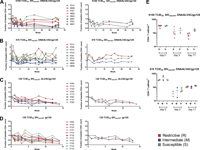
Fig. 5.
CD4+ T cell count in blood and rectal mucosa in vaccinated rhesus macaques. (A) Fraction of baseline of CD4+ T cell in the blood from eight animals challenged with 6,100 TCID50 of SIVmac251 and vaccinated with DNA/ALVAC/gp120 in the left panel. The right panel shows the averages of five R, one M, and two S animals. (B) The relative CD4+ T cell counts of nine animals challenged with 470 are depicted in the left panels, whereas the averages of the two R, three M, and four S animals are shown in the right panels. Nineteen animals were infected with 120 TCID50 of SIVmac251; eight of them were vaccinated with ALVAC/gp120 (C), and eleven were vaccinated with gp120 only (D). The averages of the R, M, and S animals from these two groups are shown in the right panels. (E) The CD4+ T cell count was evaluated in the rectal mucosa at 0, 3, and 11 weeks postchallenge in animals vaccinated with DNA/ALVAC/gp120 and exposed to 6,100 (upper panel) and 470 TCID50 of SIVmac251 (lower panel).

