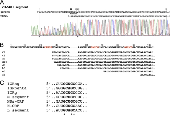
Fig. 5.
Mapping of the 3′ end of the L mRNA. (A) 3′ RACE analysis of L mRNA from ZH548-infected cells collected at 10 h p.i. The 5′ noncoding sequence of the L segment is shown in the genomic sense from positions 1 to 90. The chromatogram of the RT-PCR product corresponding to the 3′ ends of the L antigenome/mRNA is presented. The initial position of the internal in vitro polyadenylation sequence is indicated by an arrow at position 28. (B) Sequence obtained from individual PCR products after cloning into pCRII-Topo plasmids. The sequences of the antigenome were obtained from 37 plasmids. The poly(A) added in vitro is not shown. The sequences highlighted in red indicate possible transcription termination motifs. (C) Alignment of the sequences surrounding the transcription termination site in the IGR of the genome (g), antigenome (ag), N, NSs, M, and L ORFs.

