Fig. 1.
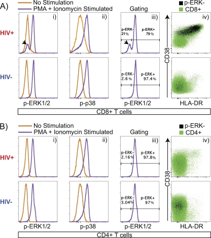
Identification of a CD8+ T cell subset refractory to PMA+I-induced ERK1/2 phosphorylation. (A) (Panel i) CD8+ T cells exhibit a shift in phosphorylation-specific ERK1/2 MFI (p-ERK1/2) in response to PMA+I stimulation. A subset of CD8+ cells that fails to phosphorylate ERK1/2 (black arrowhead) is expanded during HIV-1 infection. (Panel ii) CD8+ T cells exhibit a shift in phosphorylation-specific p38 MFI (p-p38) in response to PMA+I stimulation. (Panel iii) CD8+ T cells were gated for CD8+ p-ERK1/2-refractory (p-ERK−, black arrowhead) and p-ERK1/2-responsive (p-ERK+) subsets. (Panel iv) CD8+ T cells exhibit a p-ERK− subset. p-ERK1/2-refractory cells are predominantly activated (CD38+ HLA-DR+) and are increased in frequency during early untreated HIV-1 infection. (B) (Panel i) CD4+ T cells exhibit a shift in p-ERK1/2 MFI in response to PMA+I stimulation. (Panel ii) CD4+ T cells exhibit a shift in p-p38 MFI in response to PMA+I stimulation. (Panel iii) CD4+ T cells exhibit gating for CD4+ p-ERK1/2-refractory (p-ERK−) and -responsive (p-ERK+) populations. (Panel iv) CD4+ T cells exhibit a CD4+ p-ERK− population. Data are from one HIV− patient and one HIV+ patient.

