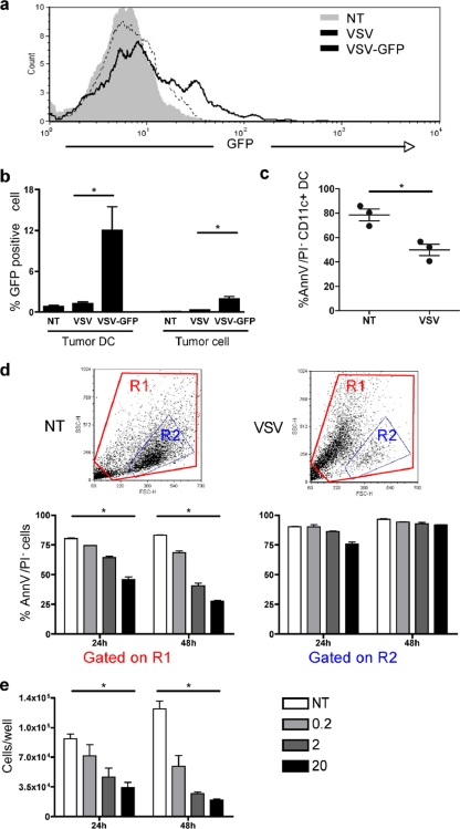
Fig. 5.
VSV infection of tumor dendritic cells reduces viability. (a and b) B16 tumors were either left untreated or injected with parental VSV or VSV-GFP. (a) The GFP expression level in CD11c+ tumor DC was analyzed by FACS at 10 h postinjection. (b) The percentage of tumor DC or tumor cells expressing GFP was also analyzed (n = 3). Parental VSV without GFP was used as negative control to account for autofluorescence induced upon virus infection. (c) The viability of CD11c+ tumor DC was assessed in B16 tumors at 10 h after VSV injection by annexin V/PI staining (n = 3). (d and e) BMDC were infected with VSV-GFP at an MOI of 0.2, 2, or 20, and cell viability was monitored 24 h and 48 h later. (d) FSC/SSC dot plots at 48 h after infection with VSV (MOI of 20) or noninfected (NT). Cell death was assessed using annexin V/PI and analyzed based on region R1 (all events) or region R2 as previously reported (1, 5). (e) Cell death was assessed by live cell counting using trypan blue (n = 3). *, P < 0.05.

