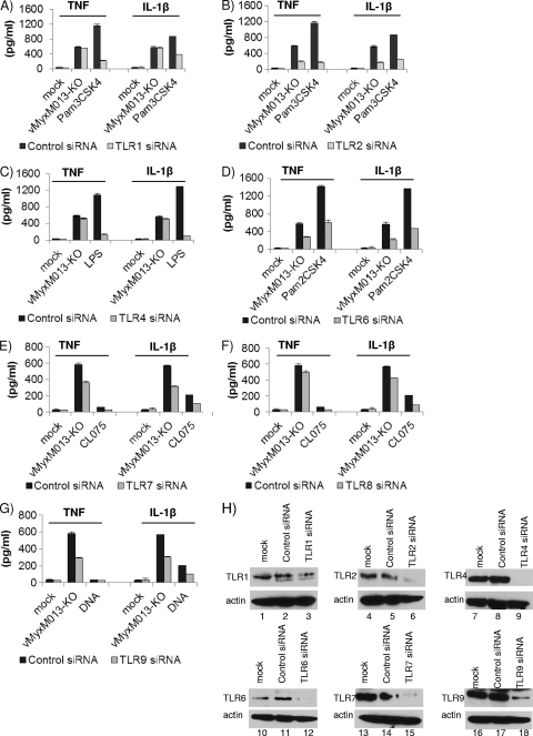
Fig. 6.
Roles of TLRs in the activation of inflammasomes and NF-κB signaling by vMyxM013-KO. THP-1 cells were transfected with siRNAs for nontargeting control or human TLRs (1 to 10), differentiated for 12 to 18 h with PMA, and then infected with vMyxM013-KO virus (MOI, 3) or treated with known ligands for TLRs: LPS (100 ng/ml), Pam3CSK4 (10 ng/ml), Pam2CSK4 (0.1 ng/ml), CL075(5 μg/ml), or E. coli DNA (6 μg/ml; control). Cell supernatants were collected after 6 h of infection or treatment with LPS, Pam3CSK4, or Pam2CSK4 and 24 h after treatment with CL075 and E. coli DNA to measure the secretion of TNF and IL-1β by ELISA. Results shown here are from A) TLR1 (A), TLR2 (B), TLR4 (C), TLR6 (D), TLR7 (E), TLR8 (F), or TLR9 (G) siRNA-treated THP-1 cells. Data are means ± SD of triplicate samples from one experiment and are representative of two or three independent experiments. P was <0.05 under all conditions. (H) Western blots showing the levels of protein knockdown when using TLR1, TLR2, TLR4, TLR6, TLR7, and TLR9 siRNAs.

