
Fig. 2.
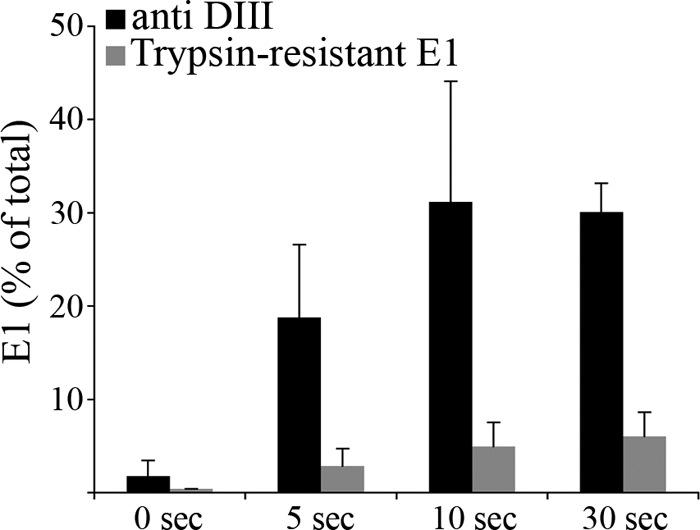
The target for exogenous domain III binding is formed at an early stage of the fusion reaction. BHK cells were incubated with radiolabeled SFV for 90 min on ice. The cells and bound virus were then treated for the indicated times on ice at pH 5.5 in the presence of 2 μM SFV DIII. The cells were washed at neutral pH and lysed. Aliquots of the lysates were immunoprecipitated with antibody to the hexahistidine tag on DIII or with polyclonal antiserum against the E1-E2 proteins, and the retrieved E1 was quantitated by SDS-PAGE and phosphorimaging. The amount of E1 retrieved by exogenous DIII is expressed as a percentage of the total amount of E1 retrieved with the polyclonal serum (black bars). The formation of trypsin-resistant E1 (gray bars) was determined as described in Materials and Methods, using parallel samples treated for the indicated times on ice at pH 5.5 in the absence of exogenous DIII. Data shown represent the mean and standard deviation of the results of 4 independent experiments (5-, 10-, and 30-s time points) or the average and range of the results of 2 independent experiments (0-s time point).