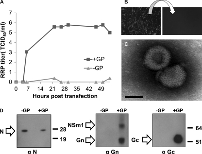
Fig. 9.
Characterization of RRPs. (A) BHK-Rep cells were either left untreated (−GP) or transfected with pCAGGS-M (+GP), and RRP titers in the collected supernatant were determined at different time points posttransfection. (B) To demonstrate that RRPs are nonspreading, BHK cells were infected with RRPs, and after 2 days, eGFP expression was observed in infected cells (left). Fresh BHK cells were incubated with the collected supernatant and monitored for eGFP expression after 3 days (right). (C) Electron micrograph of RRPs. Concentrated RRPs were stained with 1% PTA and analyzed by TEM. Bar, 50 nm. (D) To visualize RRP proteins, culture medium of BHK-Rep cells (−GP) or of BHK-Rep cells transfected with pCAGGS-M (+GP) was ultracentrifuged at 100,000 × g for 2 h. The proteins present in the pellets were separated in 4 to 12% bis-Tris gels and subsequently transferred to nitrocellulose blots. Specific proteins were detected by an anti-Gn (α Gn) or anti-Gc (α Gc) peptide antiserum or a MAb specific for the N protein (α N). The positions of the NSm, Gn, Gc, and N proteins are indicated by arrows. Molecular weight standards are indicated on the right, in thousands.

