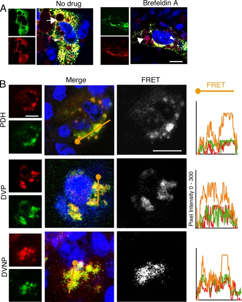
Fig. 6.
DHBV colocalizes with sphingolipid. (A) DHBV-infected PDHs were pulsed with BODIPY-TR-C5 ceramide, fixed, and stained. DHBV (green) colocalized with all species of sphingolipid (red), which persists with brefeldin A treatment. Sphingolipid accumulation in bile ducts (small arrows) and in lipid vacuoles (large arrows) can be seen. (B) Infected polarized PDHs and DVP cells and nonpolarized DVNP cells show ubiquitous virus-lipid colocalization. FRET between virus and newly synthesized sphingolipid is marked in both polarized and nonpolarized cells. The pixel intensity along a line through the colocalized regions (orange) is shown in the graphs on the right. Blue, nuclei and TOTO-3. Bar, 5 μm.

