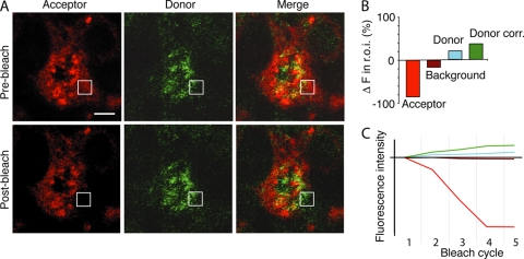
Fig. 7.
FRET between sphingolipids and virus. (A) DHBV-infected PDHs were pulsed with BODIPY-TR-C5 ceramide, fixed, and stained. The indicated square region of interest (r.o.i.) was photobleached over several cycles using a 543-nm laser line. Acceptor excitation-emission and donor excitation-emission images were then taken of the whole field. Donor fluorescence change in the r.o.i. was corrected for background photobleaching. (B) The acceptor fluorophore (red) was bleached, resulting in a quantifiable increase in the donor intensity (green) of over 15% above background in the r.o.i. (C) Progressive bleaching of the acceptor molecule over 5 cycles resulted in background bleaching (maroon), but in the r.o.i., the donor fluorophore intensity (blue) increased, resulting in a significant corrected fluorophore increase (donor correction [Donor corr.]; (green). The relative fluorescence intensity from the initial image with each bleach cycle is shown. Data shown are representative of at least 3 experiments with five regions bleached in each experiment. Bar, 5 μm.

