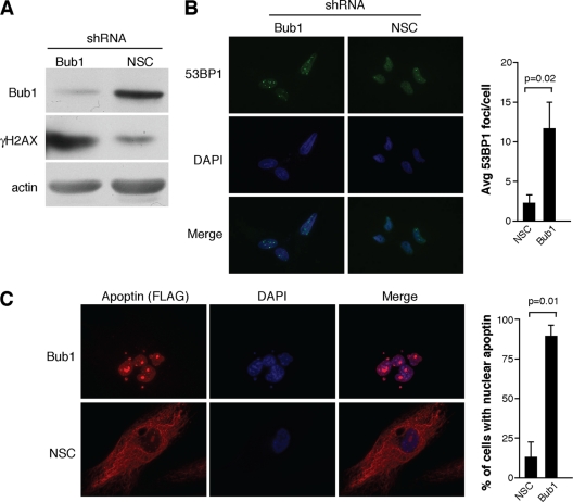
Fig. 1.
Bub1 knockdown in primary cells induces DNA damage and causes Apoptin to localize to the nucleus. (A) Immunoblot for γH2AX and bub1 from MRC5 normal fibroblasts infected with lentiviral vectors encoding an shRNA targeting bub1 or a nonsilencing control. An actin blot is included as a loading control. (B) Immunofluorescence detecting DNA damage foci in MRC5 fibroblasts treated as described for panel A. Foci were detected using an antibody against 53BP1, and cell nuclei were visualized with DAPI. Average numbers of DNA damage foci counted in knockdown and control cells are shown in the graph on the right. (C) Immunofluorescence detecting FLAG-tagged Apoptin in bub1 knockdown and control cells infected with Ad-Apwt. Cells were fixed and stained with anti-FLAG antibody, and cell nuclei were visualized with DAPI. The percentages of cells displaying nuclear localization of Apoptin are shown in the bar graph on the right. Error bars indicate standard deviations.

