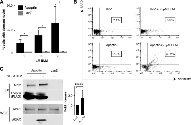
Fig. 3.
Treatment of cells with apoptin and BLM induces apoptosis in primary cells. (A) MRC5 fibroblasts were infected with Ad-Apwt or Ad-LacZ and then treated with BLM or vehicle control. Cells were fixed and stained with anti-FLAG antibody to detect Apoptin and DAPI to visualize nuclei. The morphology of the nuclei was examined by microscopy, and the percentage of cells with aberrant nuclei was determined. *, P < 0.05. (B) MRC5 fibroblasts were treated as described for panel A, stained with AnnexinV and 7AAD, and analyzed by flow cytometry. The percentage of Annexin-positive cells is indicated for each treatment (boxed). (C) MRC5 fibroblasts were infected with Ad-Apwt or Ad-LacZ and then treated with BLM. Apoptin was immunoprecipitated (IP) from cell extracts using an anti-FLAG antibody. IPs were resolved by SDS-PAGE, and immunoblotting was performed for APC1 and FLAG (Apoptin). Whole-cell extracts (WCE) used for IP were analyzed by Western blotting to confirm equal amounts of APC1 and to confirm activation of DDR (γH2AX). Band intensities of APC1 IPs were quantitated from 4 separate experiments and plotted as fold induction (right). Error bars indicate standard deviations.

