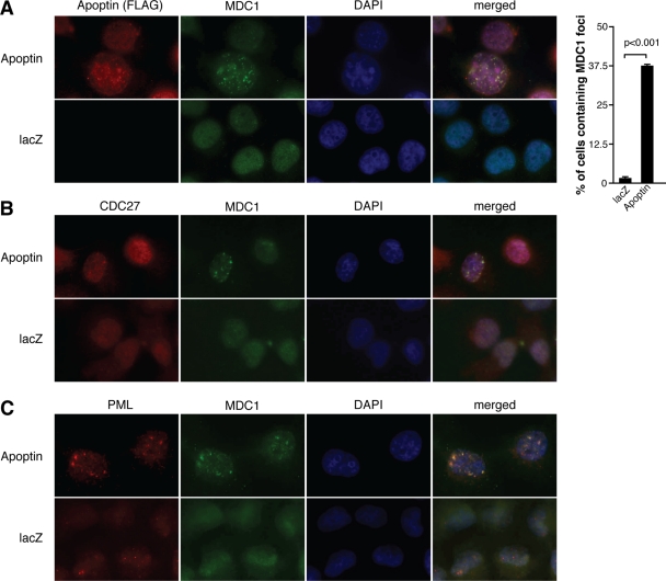
Fig. 8.
Apoptin induces MDC1 localization to PML bodies with the APC/C. H1299 cells were infected with Ad-Apwt or Ad-LacZ for 48 h and then fixed, and immunofluorescence was performed with the following antibodies: anti-FLAG (Apoptin) and anti-MDC1 antibodies (A), anti-MDC1 and anti-CDC27 antibodies (B), and anti-MDC1 and anti-PML antibodies (C). In all experiments, cell nuclei were visualized using DAPI. The percentages of cells containing MDC1 foci were determined by microscopy and are graphed on the right. Error bars indicate standard deviations.

