Fig. 7.
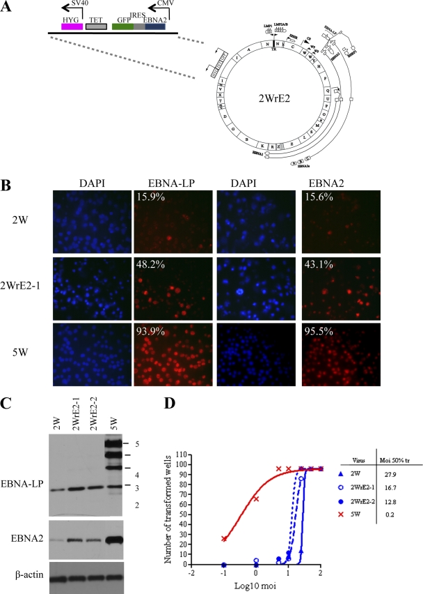
EBNA-2 repair of the 2W virus. (A) Map of the 2WrE2 BAC, including details of the EBNA2 expression cassette that was recombined into the 2W BAC genome in place of the GFP-tetracycline-hygromycin selection cassette. (B) EBV latent antigen expression in B cells infected with 2W, EBNA2-repaired 2WrE2-1, and 5W viruses. Immunofluorescence staining was performed for EBNA-LP and EBNA2 in primary B cells exposed to virus at an MOI of 50 at 3 days p.i. DAPI (blue) staining shows all nuclei in the field. The percentages of cells positive for EBNA-LP and EBNA2 (red) staining are shown in the corners of the images. (C) EBNA-LP and EBNA2 expression in 2WrE2-infected cells. Primary B cells were infected with 2WrE2 virus preparations derived from 2 different 293 producer clones (2WrE2-1 and -2) or 2W and 5W control viruses at an MOI of 50. Cell lysates at 3 days postinfection were subjected to Western blotting to determine levels of EBNA-LP and EBNA2 expression, with β-actin serving as a loading control. (D) Transformation efficiency of the EBNA2-repaired virus. Transformation assays were carried out with 2WrE2-1 and -2 or with 2W and 5W control viruses (as described for Fig. 1) to determine the efficiency of transformation, expressed as the MOI required for 50% transformation.

