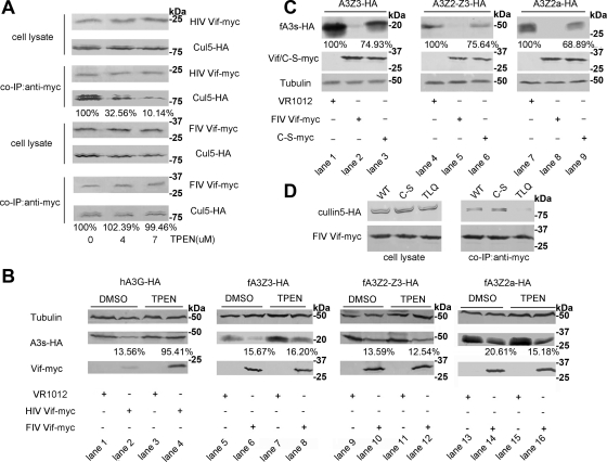
Fig. 5.
FIV Vif selects Cul5 via a novel mechanism. (A) 293T cells (106) were cotransfected with 1 μg of VR-HIV Vif-myc or VR-FIV Vif-myc and 1 μg of VR-Cul5-HA. Cells were treated with increasing concentrations of TPEN (0, 4, and 7 μM) or control DMSO 12 h before harvesting. The cell lysates were coimmunoprecipitated and analyzed by Western blotting with anti-HA and anti-myc antibodies. The percentages of Cul5 binding to Vif with different concentrations of TPEN were calculated relative to that in the absence of TPEN (set to 100%). (B) 293T cells (106) were cotransfected with 100 ng of PC-hA3G-HA, VR-fA3Z3-HA, VR-fA3Z2-Z3-HA, or VR-fA3Z2a-HA and 400 ng of VR-HIV Vif-myc, VR-FIV Vif-myc, or VR1012. Transfected cells were treated with the membrane-permeable zinc chelator TPEN at 7 μM (lanes 3, 4, 7, 8, 11, 12, 15, and 16) or DMSO (lanes 1, 2, 5, 6, 9, 10, 13, and 14) at 36 h after transfection. Cells were harvested 12 h later (48 h after transfection) and then analyzed by Western blotting with anti-HA, anti-myc, and anti-tubulin antibodies. The percentages of APOBEC3s in the presence of Vif with DMSO or TPEN treatment were calculated relative to that in the absence of Vif (set to 100%). (C) CrFK cells (106) were cotransfected with 100 ng of VR-fA3Z3-HA, VR-fA3Z2-Z3-HA, or VR-fA3Z2a-HA and 400 ng of VR-FIV Vif-myc, VR-FIV Vif C-S-myc or VR1012. The stabilities of fA3 proteins were analyzed by Western blotting with anti-HA, anti-myc, and anti-tubulin antibodies. The percentages of fA3s in the presence of FIV Vif C-S mutant were calculated relative to that in the absence of FIV Vif (set to 100%). (D) 293T cells (106) were cotransfected with 1 μg of VR-Cul5-HA and 1 μg of VR-FIV Vif-myc, VR-FIV Vif C-S-myc, or VR-FIV Vif TLQ-myc. Cell lysates were precipitated with anti-myc antibody, followed by Western blotting with anti-myc and anti-HA antibodies.

