Figure 5.
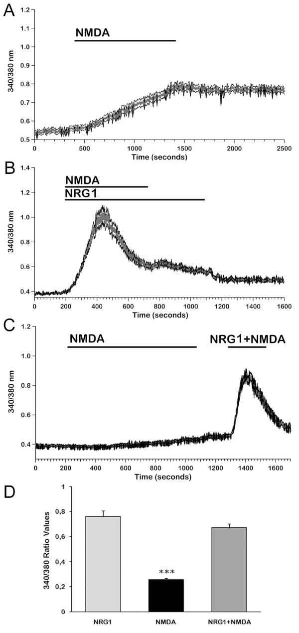
[Ca2+]i responses of ErbB4-transfected cells to NMDA and NMDA+NRG1. We measured [Ca2+]i signals induced after an acute stimulation with 8 μM NMDA and we compared them with the ones obtained co-stimulating ErbB4-transfected cells with NMDA and NRG1. A: Increases in [Ca2+]i induced by 8 μM NMDA in the presence of 2 mM extracellular [Ca2+] differ from those elicited when NRG1 is perfused: NMDA induced a very slow rise in [Ca2+]i and the plateau was reached after several minutes. Mean ± SE from a representative experiment (n = 69). B: The increase in [Ca2+]i induced by co-stimulation with NRG1 and NMDA showed the same amplitude and time course as NRG1-induced signals. Removal of NMDA slightly affected the plateau phase. Mean ± SE from a representative experiment (n = 82). C: When NRG1 was added during the slow rising response to NMDA, a standard biphasic increase in [Ca2+]i could be observed showing the same mean amplitude as with NRG1 alone. Mean ± SE from a representative experiment (n = 90). D: Histogram representing mean values of maximum amplitude (ΔR) responses to NMDA, NRG1 and NMDA+NRG1 in Tyrode Standard solution. (n = 197 with NRG; 118 with NMDA and 216 with NMDA+NRG1). Data represent means + SE from three independent experiments. *** p < 0.001 vs NRG1 and NRG1+NMDA.

