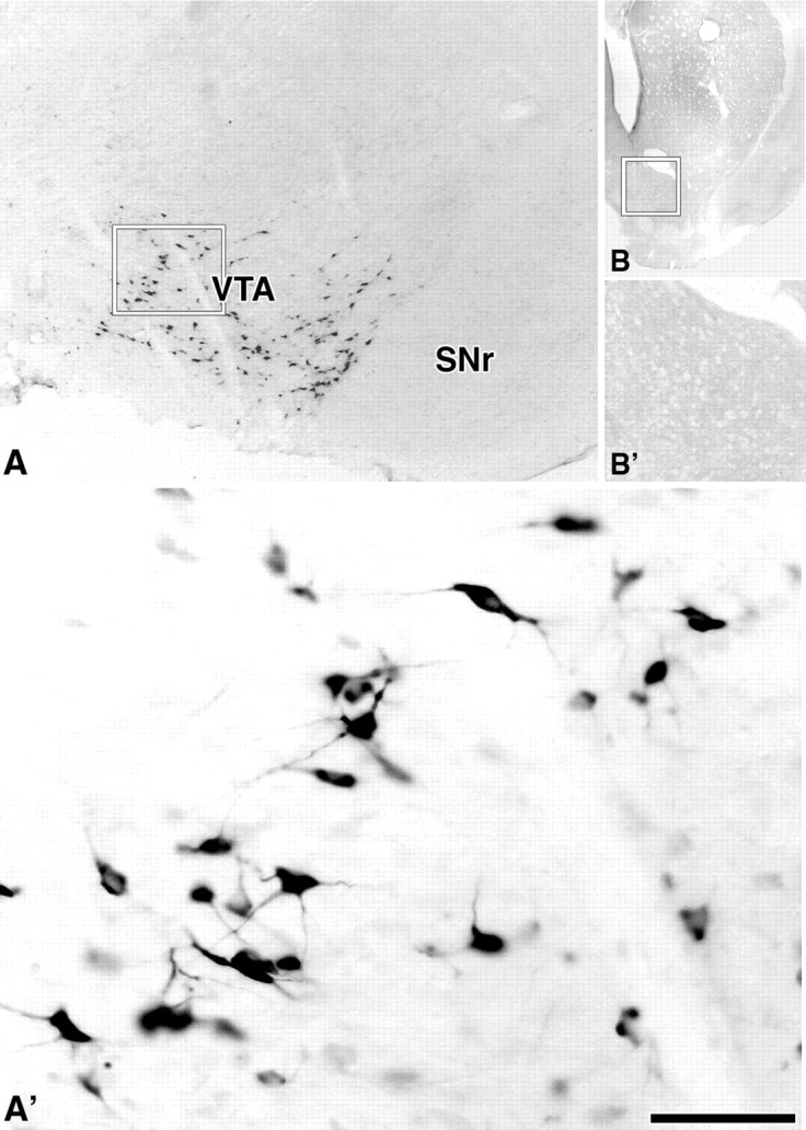
Figure 6.

Detection of WGA in VTA neurons. A, A′, WGA-immunoreactive neurons restricted to the VTA. Rectangle in A delimitates low-magnification area shown at higher magnification in A′. B, B′, Ipsilateral striatum to the VTA shown in A and A′. Rectangle in B delimitates nucleus accumbens shown at higher magnification in B′; note lack of WGA-positive fibers. SNr, Substantia nigra reticulata. Scale bar (in A′): 580 μm for A; 75 μm for A′; 2.5 mm for B; 925 μm for B′.
