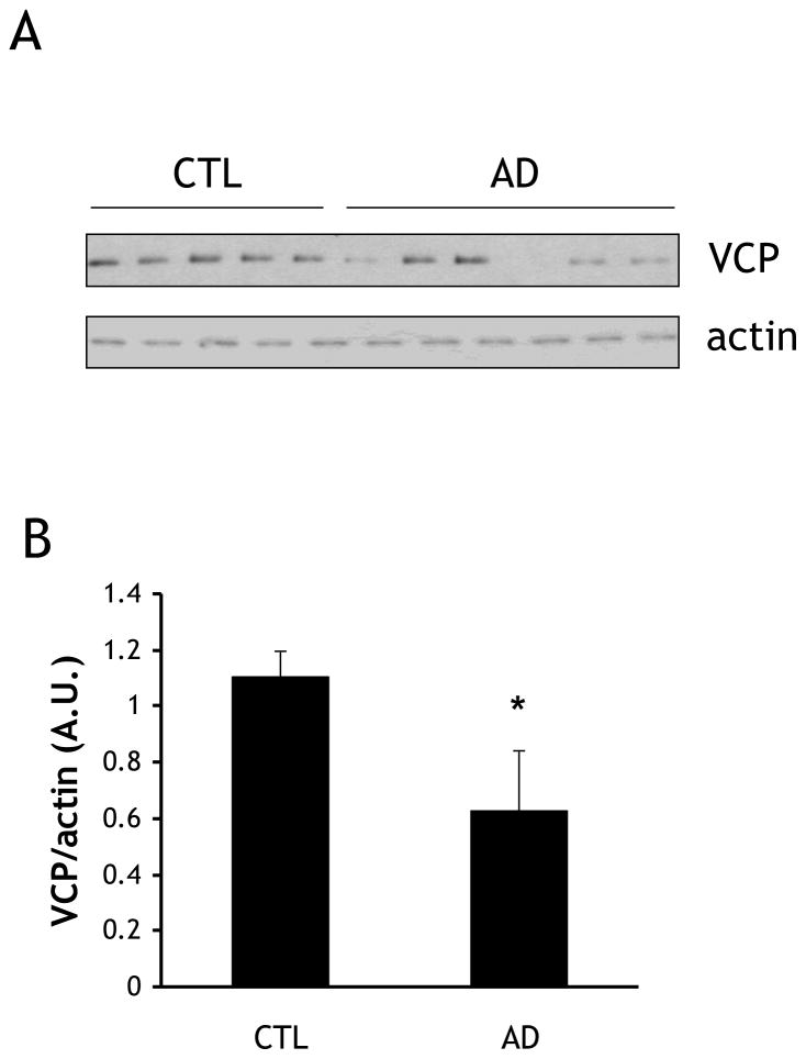
Fig. 1. VCP levels are lower in AD brain cortex than in age-matched controls.
(A) Frozen tissue from five control and six AD patients were homogenized and fractionated, and equal amounts of homogenate were immunoblotted for VCP and actin. (B) Quantification of VCP immunoblotting described in (A). Protein levels were quantified by scanning densitometry, normalized to actin, and expressed as arbitrary units (A.U.). *=p<0.05.

