
Figure 4.
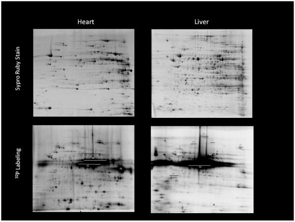
Protein content (upper panels) and autoradiograph (lower panels) of isolated heart and liver mitochondria. Mitochondria were exposed to 20 minutes of 32P labeling and then extracted identically. Protein identification and method details found in Aponte et al (Aponte et al. 2009b). Some key identifications: 12: PDH, 25:SCS, 9: β subunit Complex V, 1: aconitase