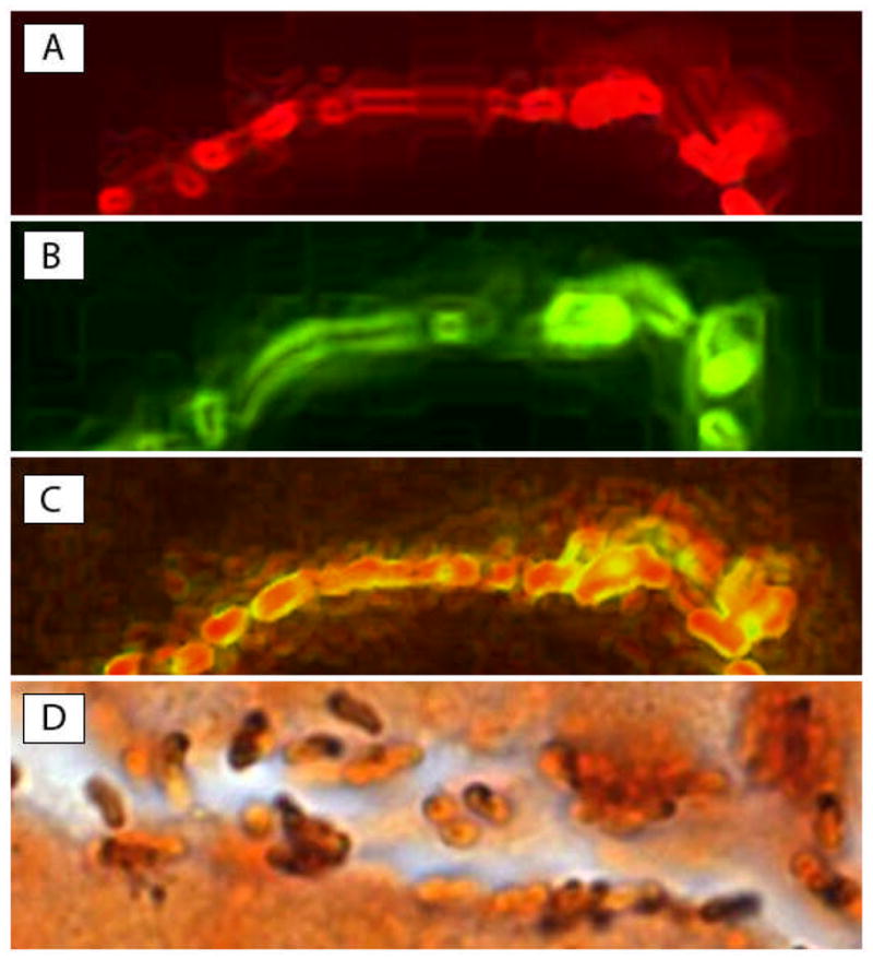
Figure 1. H. pylori in antral gastric gland.

H. pylori colonization in superficial gland of a child with duodenal ulcer in two 5-μm-thick serial sections (Original magnification X1000). (A) 16S rRNA in red (FISH-Texas red); (B) cagA in green (FISH-FITC); (C) merge of A and B, demonstrating colocalization of 16S rRNA and cagA (in yellow) in the same bacteria. Note that only cagA (green) is expressed in the outer part of bacteria while both 16S rRNA and cagA are co-localized in the center (yellow); (D) Genta stain of a gastric gland, showing many curved H. pylori-shaped bacteria colonizing the lumen mucus or adherent to the apical part of mucus-secreting cells. Note that some of the bacteria appear intensely stained black by silver, while others are weakly stained (in part or completely). These variations may represent different capture of silver stain and may be due to the presence of mucus on part of the bacterial membrane.
