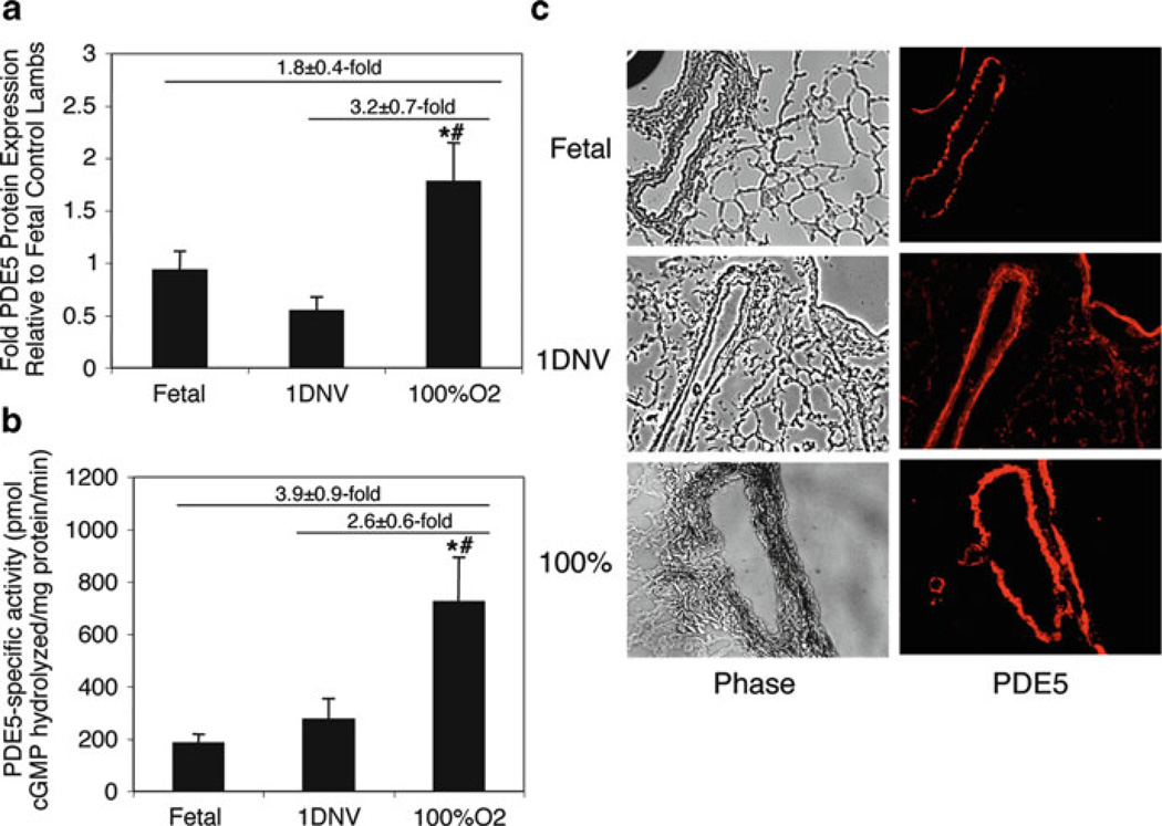
Fig. 2.
Ventilation of healthy neonatal sheep with 100% O2 induces PDE5 protein expression and activity. Ovine lung parenchyma and resistance PAs from healthy lambs ventilated with 100% O2 were harvested after 24 h and compared with both fetal lambs (Fetus) and healthy 1-day nonventilated lambs (1DNV). (a) PA PDE5 protein expression was analyzed via Western blot, with β-actin normalization. Data are shown as mean ± SEM. *P < 0.05 vs. fetal lambs, #P < 0.05 vs. 1-day nonventilated lambs. (b) Lung PDE5-specific activity was measured as the sildenafil-inhibitable fraction of total cGMP hydrolysis, normalized for total milligrams of protein (200 µM cGMP substrate in assay; 100 nM sildenafil for inhibition). Data are shown as mean ± SEM. *P < 0.05 vs. fetal lambs, #P < 0.05 vs. 1-day nonventilated lambs. (c) PDE5 expression is localized within the PA smooth muscle and nearby airways. The left column shows phase-contrast images (×10) of frozen lamb lung sections. The right column shows the corresponding sections stained for PDE5, with immunofluorescence shown as red. Reproduced with permission from Farrow et al. (2008a)

