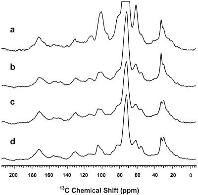
Figure 4.
Solid-state 13C-NMR spectra showing the progressive purification of the suberin from the wound-healing periderm of potato tuber slices. a, Cold-water-washed periderm preparation showing resonances from suberin, wax, and cell wall components. b, Periderm preparation treated with cellulase and pectinase, drastically reducing carbohydrate peaks centered at 72 ppm. c, Suberin fraction after exhaustive extraction of the wax with organic solvents, reducing mainly the methylene peaks at 33 ppm. d, Suberin fraction after dioxane:water extraction to remove residual sugars and soluble lignins, resulting in a better definition of the carbohydrate peaks. The glycerol resonances expected at 66 and 75 ppm were obscured by large cell wall peaks near 72 ppm and possibly by signals from suberin esters of primary alcohols at 65 ppm.

