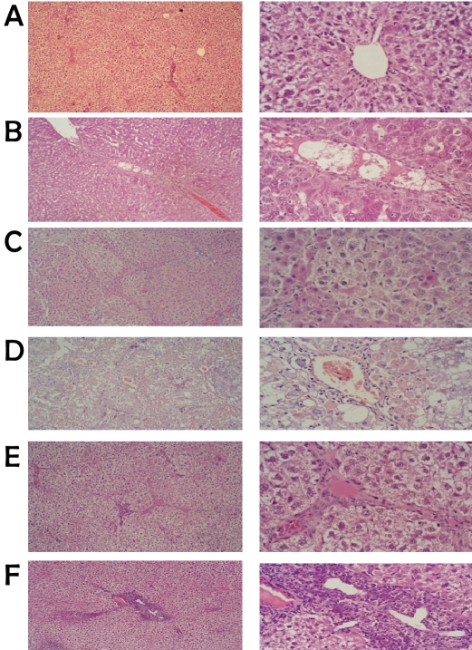
Figure 1.
Representative microscopic photographs of control versus TAA-induced rats by H & E staining (low-power on the left and high-power on the right). a) mock-treated showing normal hepatocytes. b) TAA-induction for 8 weeks, c) 16 weeks showed small nodules with degenerative hepatocytes, absence of sinusoid, an increase of fibrous tissue thickness septa, expansion of portal tract with hepatic central vein and hepatic nodules increase (cirrhosis). d) After 20 weeks of TAA induction the results showed marked macro vesicular steatosis changes of hepatocytes as well as congested sinusoidal structures and complete cirrhosis. e) Rat liver paraffin section after 2 weeks of MNCs treatment showing decrease in fibrotic thickness septa and appearance of apoptotic cells (shrinkage and dark color nucleus). f) Photograph of rat liver paraffin section after 4 weeks of MNCs treatment showing the appearance of discontinuous septa, presence of apoptotic cells and liver return to normal architecture.

