
Figure 2.
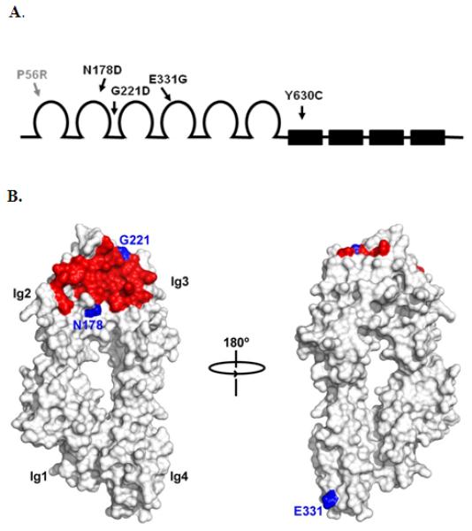
Schematic of domains of CNTN4 and approximate sites of variant amino acids. (A) The 6 Ig-like domains of CNTN4 are shown as open loops and the 4 fibronectin II-like domains as filled boxes. The sites of the variant amino acids are shown as gray (control) or black (ASD patients) arrows. (B) Surface representations of the 4 Ig-like domains of mouse CNTN4 (CNTN4Ig1-4) with binding site of PTPRG shown in red. The altered amino acids in the ASD patients within the Ig-like domains are shown in blue. The variant Y630C is not shown since this mutation lies within the FNIII region of CNTN4, the structure of which has not yet been determined.