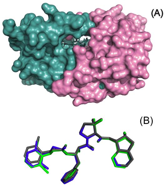
Figure 5.
The inhibitors bind to the HIV-1 protease in a similar conformation and interact with the same residues in the protease. Shown are surface representations of HIV-1 protease pseudo-wild type bound to KNI-10265 (blue), KNI-10074 (green), and KNI-10006 (gray) (Panel A). In panel B the structure of the bound compounds have been superimposed to indicate that the bind to the protease in the same extended conformation.

