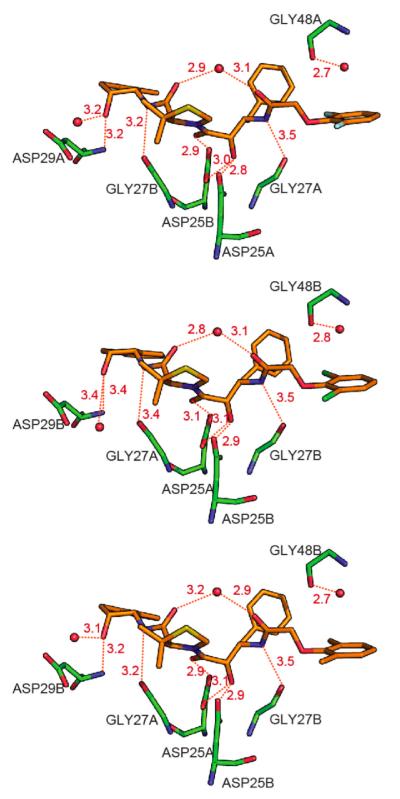
Figure 6.
Hydrogen bonding interactions between HIV-1 protease pseudo-wild type (pWT) and KNI-10265 (top), KNI-10074 (middle), and KNI-10006 (bottom). Red spheres indicate water molecules. The compound and the protein are colored by atom type with oxygen in red, nitrogen in blue, sulfur in yellow, fluorine in cyan, chlorine in green, and carbon in orange for the compound and green for the protein. Hydrogen bonds are represented by red dashed lines, and the hydrogen bond distances shown in this figure are the average value of orientation A and B. The hydrogen bond pattern is the same for the three inhibitors.

