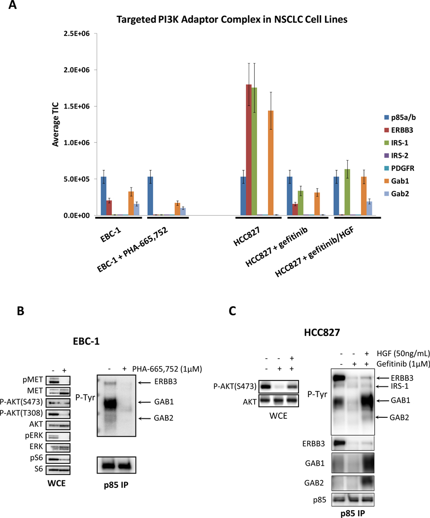
Figure 1.
(A) The quantitative output of the TIMM approach in EBC-1 and HCC827cells subjected to the indicated conditions. Cells were treated with the indicated drugs and ligands for 6 hours before lysis. The relative signal level of each detected adaptor (normalized for p85 levels) is shown. (B) EBC-1 cells were treated in the absence or presence of the MET inhibitor, PHA-665,752 (1µM) for 6 hours and then subjected to lysis. (C) HCC827 cells were treated in the absence or presence of gefitinib (1µM) and HGF (50 ng/mL) for 6 hours. Left) Whole cell extracts were probed with the indicated antibodies. Right) Extracts were subjected to a p85 IP, and the IP was probed with an anti-PTyr and anti-p85 antibodies.

