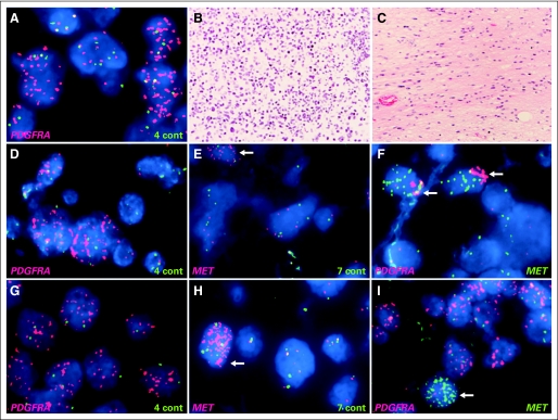
Fig 2.
Heterogeneity of focal amplification in diffuse intrinsic pontine glioma. (A) Fluorescent in situ hybridization showed high-level amplification of PDGFRA in focal area of tumor (PDGFRA, red; control chromosome 4, green). Focus of tumor with PDGFRA amplification and remainder of tumor lacking high-level PDGFRA amplification (data not shown) demonstrated similar histopathologic features; both consisted of densely packed tumor cells with minimal normal tissue. PDGFRA amplification was found in both solid groups of tumor cells (B: hematoxylin and eosin [HE] staining) and scattered infiltrating tumor cells (C: HE). Coamplification of PDGFRA and MET in BSG009T (D to F) and BSG037T (G to I). Cells with amplified PDGFRA (D, G: PDGFRA, red; control, green) and MET (E, H: MET, red; control, green) showed coamplification (F, I: PDGFRA, red; MET, green) of (F) both genes in same tumor cells (arrows) or (I) independent amplification in different tumor subclones (arrows).

