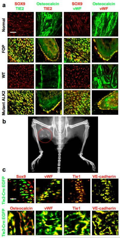
Figure 1.
Endothelial ceΠ differentiation in heterotopic ossification. (a) Immunohistochemistry of chondrogenic (first and third columns from left) and osteogenic (second and forth columns from left) lesions from Fibrodysplasia Ossificans Progressiva (FOP) patients with activating ALK2 mutations and from Cre-dependent constitutively active ALK2 (caALK2) transgenic mice. Chondrogenic lesions show co-expression of the endothelial markers TIE2 and vWF with the chondrocyte marker SOX9. Osteogenic lesions show co-expression of TIE2 and vWF with the osteoblast marker osteocalcin. Normal cartilage and bone from human hip joint (top row) or wild-type (WT) mouse knee joint (third row) show no evidence of TIE2- or vWF-positive chondrocytes or osteoblasts. Scale bar, 40 μm. (b) X-ray image of heterotopic ossification (red circle) in a Cre-induced caALK2 transgenic mouse. (c) Immunohistochemistry of BMP4-induced heterotopic cartilage and bone in Tie2-Cre reporter mice showing EGFP positive chondrocytes (top row) and osteoblasts (bottom row). These EGFP positive cells show expression of endothelial markers vWF, Tie1, and VE-cadherin. Scale bar, 20 μm.

