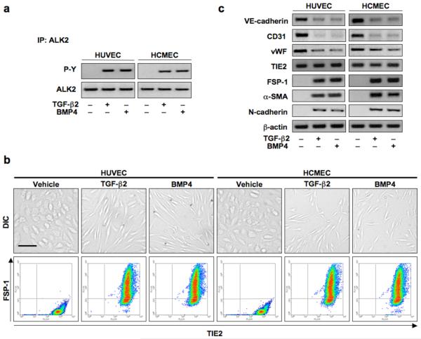
Figure 4.
TGF-β2 and BMP4 activate ALK2 and induce endothelial-mesenchymal transition. (a) Immunoblotting of immunoprecipitates confirming phosphorylation of ALK2 by 15 min of TGF-β2 or BMP4 stimulation. (b) DIC imaging, immunocytochemistry and flow cytometry showing a change in cell morphology and co-expression of TIE2 and FSP-1 in endothelial cells treated with TGF-β2 or BMP4 for 48 h. Scale bar, 20 μm. (c) Immunoblotting confirming EndMT with decreased expression of VE-cadherin, CD31, and vWF and increased expression of FSP-1, α-SMA, and N-cadherin in cells treated with TGF-β2 or BMP4. TIE2 levels remained constant.

