Figure 1.
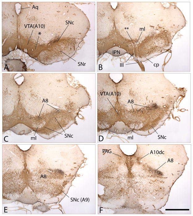
Figure 1A–F. Photomicrographs illustrating a series of sections through the mesencephalon of the rat shown from A to F in rostrocaudal order. The sections were processed to exhibit immunoreactivity against tyrosine hydroxylase, which marks ventral mesencephalic dopaminergic neurons and axons brown, and thus shows the ventral tegmental area (VTA(A10)), substantial nigra pars compacta (SNc(A9)), and retrorubral field (A8). The juncture of VTA(A10) and SNc(A9) is indicated by * in panel A, as is the zone where VTA(A10) becomes A8 by ** in panel B and continuities between A10, A9 and A8 can be observed in all of the panels. Note in panel A that the dendrites of SNc(A9) dopaminergic neurons extend downward into the substantia nigra pars reticulata (SNr). Panel F illustrates the caudomedial extension of A8 and the tyrosine hydroxylase-immunoreactive (possibly L-DOPA containing) neurons in the periaqueductal gray (PAG), which are designated as A10dc. The black substance in all of the panels marks axons projecting from the central extended amygdala, specifically from the bed nucleus of stria terminalis, that were labeled in the laboratory with a dye. Note that the labeled pathway skirts past the SNc in panels A and B to terminate relatively exclusively within A8. The labeled axons in A8 and A10dc shown panel F are enlarged in Figs. 2A and B, respectively. Other abbreviations: III - oculomotor (3rd cranial) nerve and roots; Aq - cerebral aqueduct; cp - cerebral peduncle; IPN -interpeduncular nucleus; ml - medial lemniscus. Scale bar: 1 mm.

