Figure 2.
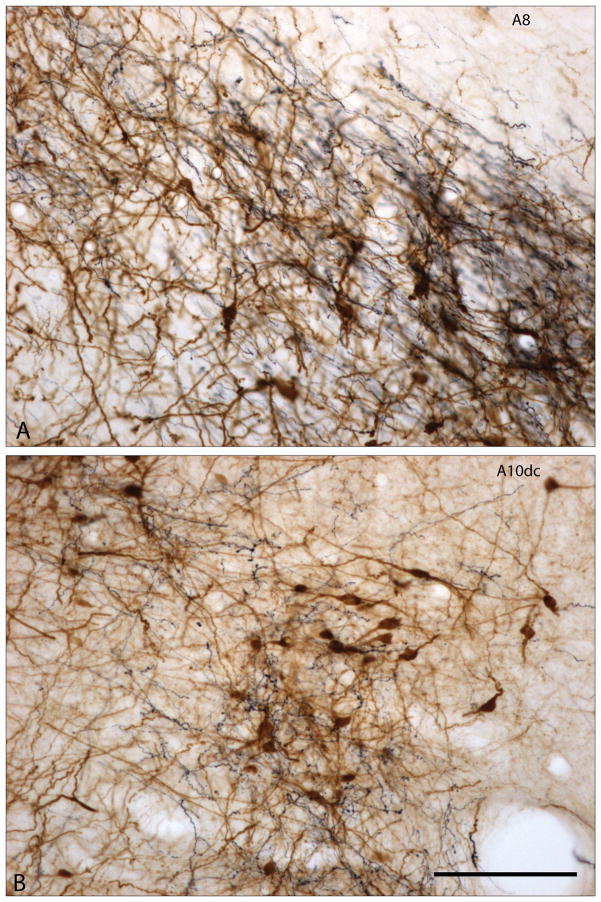
Figure 2A and B. Photomicrographs showing enlargements of areas designated as A8 and A10dc in Fig. 1F. Tyrosine hydroxylase immunoreactive elements are brown and dye-labeled axons projecting from the bed nucleus of stria terminalis are black. The dye-labeled axons form a dense plexus of fibers containing many varicosities suggestive of abundant synaptic contacts. Scale bar: 100 μm.

