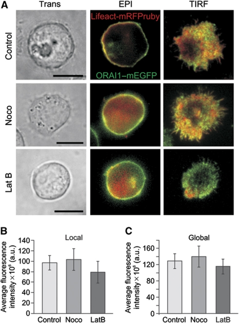
Figure 7.
Cytoskeleton polymerization-disrupting drugs do not inhibit ORAI accumulation at the IS. (A) Transmission light (Trans), epifluorescence (Epi) and TIRFM pictures of representative single Jurkat T cells treated with cytoskeleton-disrupting drugs (2 μM nocodazole, 10 μg/ml latrunculin B). Cells were transiently expressing Lifeact-mRFPruby and ORAI1–mEGFP proteins. After treatment, T-cells were settled on anti-CD3 antibody-coated coverslips for 20–30 min in the presence of extracellular Ca2+ solution to induce the formation of a matured IS. Scale bars are 10 μm. Average intensity of the local (B) and global (C) fluorescence signal of ORAI1–mEGFP measured for cells pre-treated with nocodazole or latrunculin B or kept under control conditions (control n=32, nocodazole n=20, latrunculin B n=21).

