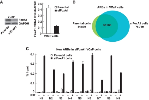
Figure 2.
FoxA1 depletion in VCaP cells. (A) FoxA1 mRNA and protein levels. VCaP cells were treated for 72 h with siRNA specific for FoxA1 mRNA (siFoxA1) or control siRNA (parental). (B) Overlap of ARBs (FDR <2%) in parental and FoxA1-depleted VCaP cells. (C) Directed ChIP validation of new ARBs in parental (white bars) and FoxA1-depleted (solid bars) VCaP cells. The cells were exposed to 100 nM DHT (+) or vehicle (−) for 2 h prior to ChIP assays. Mean+s.e.m. values for duplicate samples are shown.

