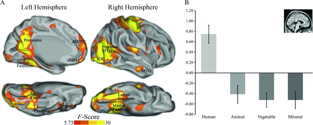
Figure 2.
(A) Brain regions showing a main effect of category from a whole-brain voxelwise repeated measures ANOVA (P < 0.05, corrected). Statistical maps of left medial hemisphere, right lateral hemisphere, and ventral surfaces of both hemispheres are overlaid onto inflated cortical renderings. (B) ROI analysis of parameter estimates in the DMPFC showed that this area was preferentially active to human social versus nonsocial scenes (t47 = 6.76, P < 0.001). Coordinates (x, y, z) are in Montreal Neurological Institute stereotaxic space. vMPFC = ventral medial prefrontal cortex.

