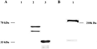
Figure 1.
A, Immunoblot analysis of the pGEX-NADH-GOGAT fusion protein. The blot of a 15% polyacrylamide gel was probed with polyclonal NADH-GOGAT antiserum and developed with alkaline-phosphatase-conjugated secondary antibodies. Lanes 1 through 3 contain 10 μg of protein each. Lane 1 contains total protein from E. coli carrying the uninduced pGEX-NADH-GOGAT construct; lane 2 contains total E. coli protein carrying the induced pGEX-NADH-GOGAT construct; and lane 3 contains the partially purified pGEX-NADH-GOGAT construct cleaved with thrombin. B, Immunoblot of total soluble nodule protein. The blot of a 6% polyacrylamide gel was probed with affinity-purified NADH-GOGAT antibodies and developed with alkaline-phosphatase-conjugated secondary antibodies. Lane 1 contains 100 μg of soluble protein of effective nodules.

